Enhancing cell adhesion on device surfaces

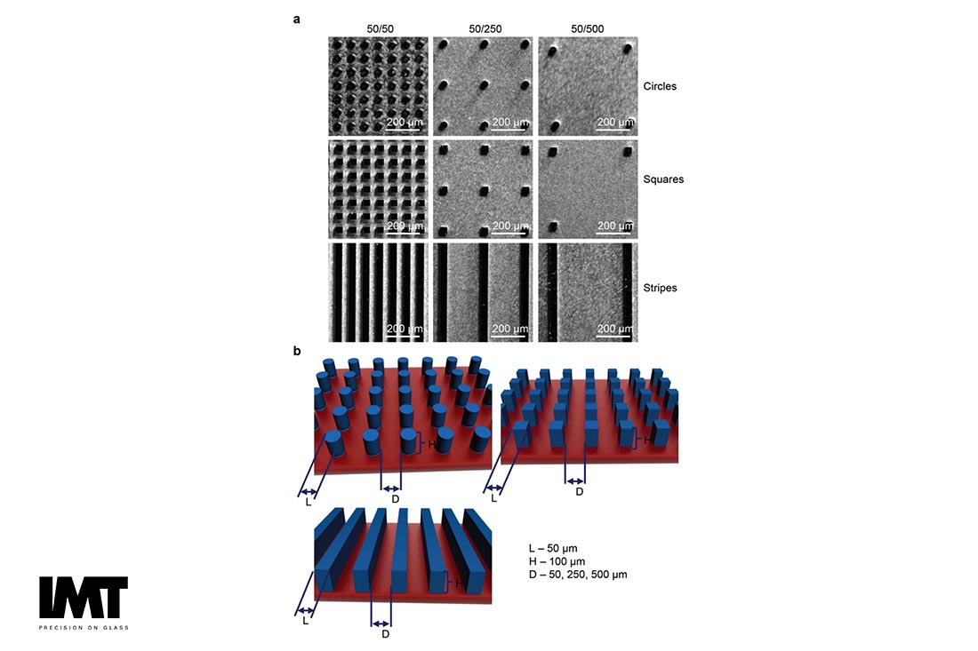
Modulation of collective cell behaviour by substrates with patterned surfaces for cell culturing. (Source: “Modulation of collective cell behaviour by geometrical constraints,” M. Lunova, V. Zablotskii, N. M. Dempsey, T. Devillers, M. Jirsa, E. Syková, Š. Kubinová, O. Lunov and A. Dejneka, Integr. Biol., 2016, 8, 1099. DOI: 10.1039/C6IB00125D -Published by The Royal
Cell adhesion is dependent on cell type, length of time on the pattern or surface, cell density, and media type. Micro and nano-patterning a surface modification can be used for enhancing cell adhesion on microdevice surfaces to confine cells to a specific region of a microfluidic flow cell. For example, Toner’s group demonstrated that collagen (cell adhesion) surrounded by albumin (non-adhesive) could create cell and cell-free zones in a chip as long as the cell culture medium was free of serum proteins. By exposing the surface to hepatocytes in serum free media, then fibroblasts in serum containing media, a two-cell type pattern of hepatocytes and fibroblasts were defined [1]. Another strategy is to use TMMF, a photostructurable material, as an intermediary layer to create so-called “2.5D” structures for cell capturing and culturing [2].
Self-assembled monolayers (SAMs) can also be used to chemically define surface functionality. Depending on the end functional group, surfaces can be tailored for adhesion, lubrication, wettability, or protein physisorption [3]. Thiol-based SAMS are the most commonly used, reacting with gold, [3], [4]. a metal readily patterned on glass. Organosilanes are a powerful chemical tool, as their reaction chemistry with the free hydroxyl groups found on the surface of the glass itself is very well-understood [4]. Common surface properties important for IVDs that can be created by organosilane modification include [3]:
- Increasing hydrophobicity for cell/biomolecule adhesion, or droplet/digital microfluidics;
- Increasing hydrophilicity to prevent biological material adsorption;
- Adding a surface charge for ionic association or other purposes.
Recently, a burst of studies utilizing cell micro- and nanopatterning techniques revealed that the manipulation of cell geometry, such as confinement to circular or square patterns independently of the cell spread area, affects many crucial cellular processes. To produce uniform substrates for biological applications, deep reactive ion etching is used to pattern silicon wafers so as to produce sets of substrates with surfaces consisting of arrays of silicon micropillars of different geometry and with different values of inter-pillar spacing. The patterned silicon substrates were coated with a parylene, which is a biocompatible, inert and very low permeability material [2], [5].
Alternative scenarios can be envisaged by the structuring of a class of photo-definable materials known as Photoimaginable Bonding Adhesives (PBA). PBAs can be photo-patterned to create complex fluidic channel systems for the capture and fusion of cells or droplets directly on a glass wafer by using standard MEMS processes [6]. This method is far more economical then typical hybrid approaches as it does not require extra passivation layers due to the availability of bio-compatible PBAs. The combination of bio-functionalization and UV-adhesive sealing can lead to a cost-effective and up-scalable production of cell-trapping flow cells [5].
Works Cited
[1] A. Folch and M. Toner, "Cellular micropatterns on biocompatible materials," Biotechnol Prog., vol. 14, no. 3, pp. 388-392, 1998.
[2] M. Lunova, V. Zablotskii, N. M. Dempsey, T. Devillers, M. Jirsa, E. Sykova, S. Kubinova, O. Lunov and A. Dejneka, "Modulation of collective cell behaviour by geometrical constraints," Integr. Biol., vol. 8, p. 1099, 2016.
[3] N. R. Glass, R. Tjeung, P. Chan, L. Y. Yeo and J. R. Friend, "Organosilane deposition for microfluidic applications," Biomicrofluidics, vol. 5, no. 036501, 2011.
[4] N. Li, A. Tourovskaia and A. Folch, "Biology on a Chip: Microfabrication for Studying the Behavior of Cultured Cells," Critical reviews in biomedical engineering, vol. 31, no. 0, pp. 423-428, 2003.
[5] A. M. Skelley, O. Kirak, H. Suh, R. Jaenisch and J. Voldman, "Microfluidic Control of Cell Pairing and Fusion," Nat Methods. , vol. 6, no. 2, pp. 147-152, 2009.
[6] U. Stöhr , P. Vulto , P. Hoppe , G. Urban and H. Reinecke, "High-resolution permanent photoresist laminate for microsystem applications," J. Micro/Nanolith. MEMS MOEMS, vol. 73, 2008.
[7] L. Wang, C. Zhu, Q. Zheng and X. He , "Preparation of Homogeneous Nanostructures in 5
Minutes for Cancer Cells Capture," Journal of Nanomaterials, vol. 2015, no. Article ID
391850, p. http://dx.doi.org/10.1155/2015/391850, 2015.

